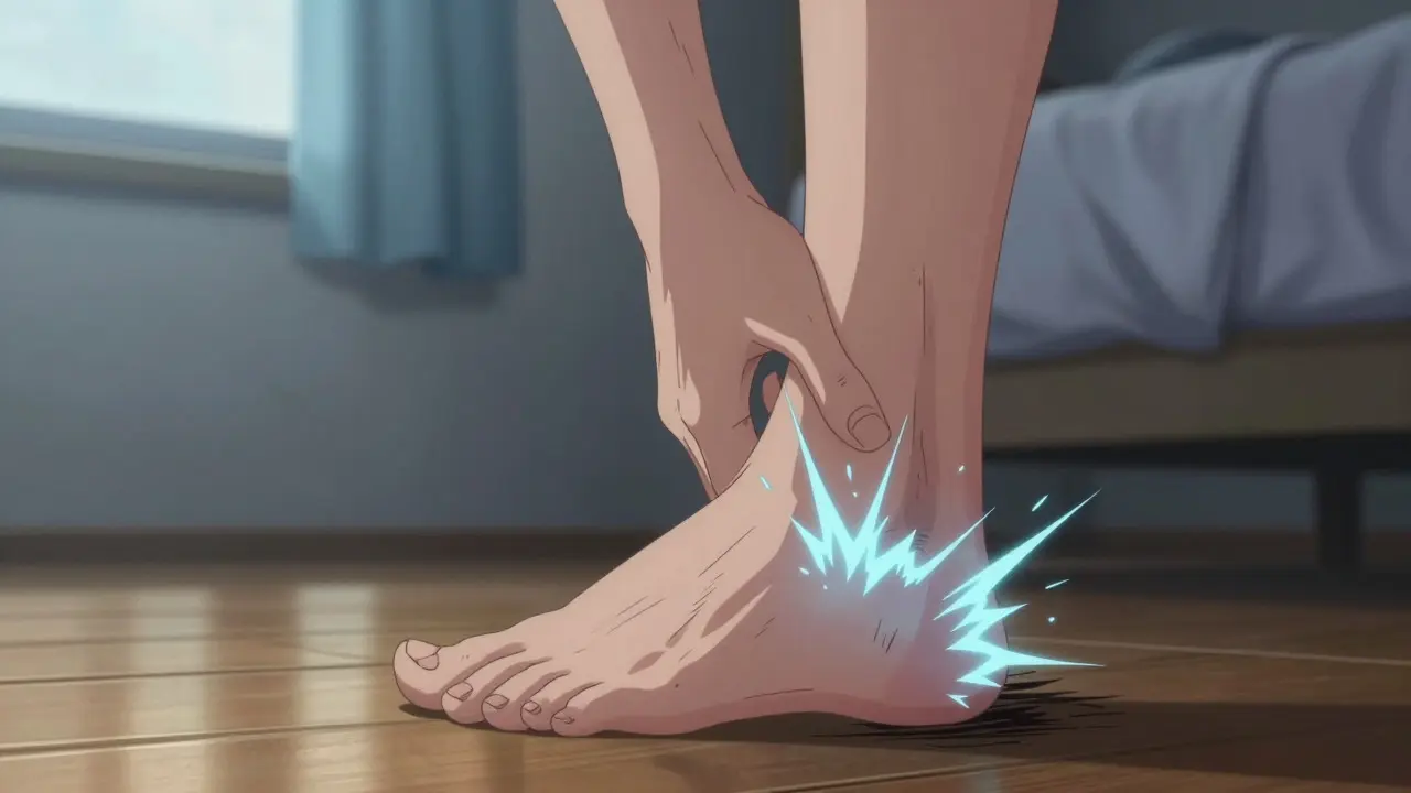
Waking up to a sharp, stabbing pain in your heel that makes your first few steps feel like walking on broken glass is a nightmare for millions of people. If you've ever limped out of bed or winced after standing at work all day, you are likely dealing with plantar fasciitis, a condition affecting the thick band of tissue connecting your heel bone to your toes. While it’s often called an inflammatory issue, recent medical consensus suggests it is actually a degenerative problem known as a biomechanical overuse condition affecting the plantar fascia. This guide breaks down why this happens, who is at risk, and which treatments actually work based on current clinical evidence.
Key Takeaways
- Plantar fasciitis affects roughly 10% of adults at some point, with peak incidence between ages 40 and 60.
- The primary symptom is sharp pain during the first steps after waking or prolonged rest, not constant throbbing.
- Conservative treatments like specific stretching and orthotics resolve 80-90% of cases within 6-12 months.
- Corticosteroid injections provide quick relief but carry risks and are less effective long-term than structured physical therapy.
- Risk factors include high BMI, limited ankle flexibility, flat feet, and jobs requiring long hours of standing.
Understanding the Root Cause: Degeneration, Not Just Inflammation
For years, doctors treated heel pain as a simple inflammation, hence the name "fasciitis." However, histologic examinations of tissue from patients undergoing surgery reveal myxoid degeneration and fragmentation rather than significant inflammation. This shift in understanding led experts to prefer the term plantar fasciopathy. The plantar fascia originates from the posteromedial calcaneal tuberosity (the heel bone) and inserts into each metatarsal head, forming the foot's longitudinal arch. When this tissue undergoes repetitive stress without adequate recovery time, micro-tears occur at its attachment point on the heel. Over time, these tears fail to heal properly, leading to structural changes and chronic pain.
This degenerative process explains why the pain is often worst in the morning. After sleeping, the fascia contracts and shortens. Your first few steps force it to stretch abruptly, triggering a painful response. As you walk, the tissue warms up and loosens, which is why the pain often subsides after 5-10 minutes of activity. However, returning to the area later in the day or after sitting for long periods can bring the pain back.
Who Is at Risk? Identifying Key Factors
Not everyone develops this condition. Certain lifestyle and anatomical factors significantly increase your odds. According to clinical guidelines from the American Academy of Family Physicians, several risk factors stand out with strong statistical correlations:
- Body Mass Index (BMI): Individuals with a BMI greater than 27 kg/m² have an odds ratio of 4.2 for developing the condition. Excess weight places additional mechanical load on the fascia.
- Ankle Flexibility: Limited ankle dorsiflexion (less than 10 degrees) increases risk by a factor of 3.7. Tight calves pull on the heel, straining the fascia.
- Foot Structure: People with pes planus (flat feet) have an odds ratio of 2.9 due to altered biomechanics and increased tension on the arch.
- Occupation: Jobs requiring more than 4 hours of daily standing, such as teaching or factory work, carry an odds ratio of 5.1.
Age also plays a role, with prevalence peaking in the 45-64 age group at 10.2%, compared to just 3.6% in those aged 18-44. Interestingly, the condition has a bimodal distribution: it affects both sedentary individuals with higher BMIs and active runners who log more than 10 miles per week.
Diagnosis: More Than Just an X-Ray
Many patients arrive at the clinic asking about "heel spurs," believing a bony growth is the culprit. Dr. James D. Heckman, former Chairman of Orthopaedics at Thomas Jefferson University, notes that 80% of patients with a clinical diagnosis of plantar fasciitis have no radiographic evidence of spurs. Conversely, 15% of asymptomatic individuals show spurs on X-rays. Therefore, an X-ray alone is insufficient for diagnosis.
Accurate diagnosis relies on clinical features established by the American Orthopaedic Foot & Ankle Society (AOFAS). A doctor will typically look for three out of four specific signs:
- Localized tenderness 2-3 cm anterior to the medial calcaneal tuberosity.
- Pain with first-step morning ambulation.
- Pain with passive dorsiflexion of the toes (pulling your toes up while seated).
- Pain with prolonged standing.
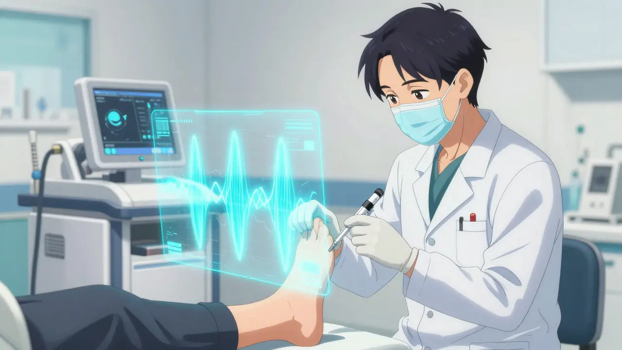
If symptoms persist, ultrasound imaging may be used. Normal plantar fascia thickness ranges from 2.0 to 3.5 mm. Ultrasound reveals thickness greater than 4.0 mm in affected patients, offering 85% sensitivity and 90% specificity for diagnosis. It is crucial to differentiate this from other conditions like Baxter neuritis (burning pain) or tarsal tunnel syndrome (numbness/tingling), which require different treatments.
First-Line Treatments: Stretching and Lifestyle Changes
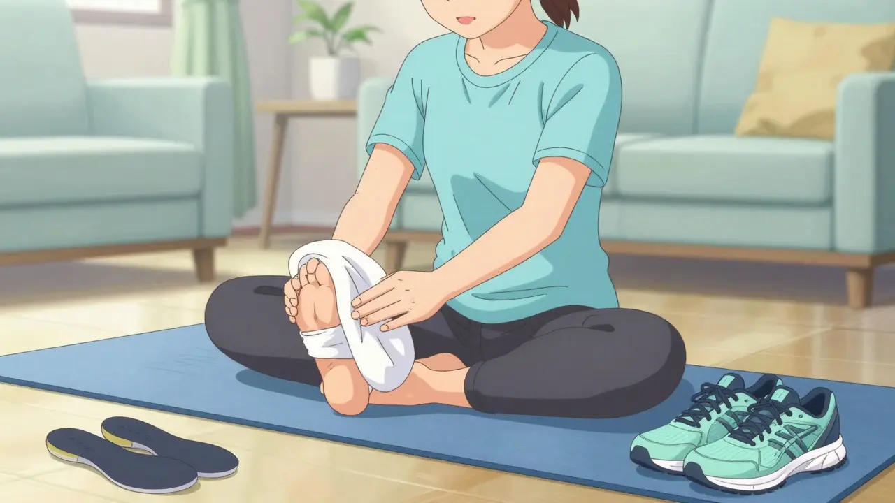
Before considering injections or surgery, the gold standard remains conservative management. The American Academy of Orthopaedic Surgeons (AAOS) strongly recommends plantar fascia-specific stretching as the first-line treatment. This is distinct from general calf stretches. Effective protocols target the fascia directly through towel stretches where the toes are maximally dorsiflexed (pulled toward the shin).
A typical routine involves:
- Sitting on the floor with legs extended.
- Wrapping a towel around the ball of your foot.
- Gently pulling the towel toward you to stretch the sole of the foot.
- Holding for 10 seconds, repeating 10 times, 3 times daily.
Consistency is key. Studies show that 92% adherence is needed for optimal results, with significant improvement seen in 8-12 weeks. Additionally, weight loss is strongly recommended for overweight patients; a reduction of just 1 point in BMI correlates with a 5.3% pain reduction at 6 months.
Supportive Devices and Physical Therapy
Stretching works best when combined with proper support. Custom orthotics demonstrate a 68% pain reduction compared to 52% for prefabricated ones at 12 weeks. When selecting shoes, look for a heel-to-toe drop of 10-15mm and solid medial arch support. Popular options among patients include models like the Brooks Adrenaline GTS and Hoka Clifton, which offer substantial cushioning and stability.
Night splints are another tool in the arsenal. These devices hold the foot in a neutral position while you sleep, preventing the fascia from contracting overnight. A randomized controlled trial found that night splints showed 72% improvement at 6 weeks versus 45% with placebo. However, user compliance is a challenge; about 44% of users discontinue use due to sleep discomfort. For those who tolerate them, wearing the device for 4-5 hours nightly can yield therapeutic benefits.
Physical therapy sessions, typically costing $80-$120 per session, can accelerate recovery. Therapists may use manual therapy, dry needling, or guided exercise programs. Medicare covers 80% of these costs after the deductible is met, making it a viable option for many.
Advanced Interventions: Injections and Shockwave Therapy
If conservative measures fail after 3 months, doctors may consider more invasive options. Corticosteroid injections are common but come with caveats. They provide rapid relief, but only temporary-average duration is 4.2 weeks. Furthermore, multiple injections carry an 18% risk of plantar fascia rupture and a 22% risk of fat pad atrophy. Consequently, guidelines recommend against early injection, yet 30-40% of patients receive them within the first 3 months of diagnosis.
Extracorporeal shockwave therapy (ESWT) is a non-invasive alternative that uses sound waves to stimulate healing. It achieves 70-80% success rates after 3-4 treatments. However, it is expensive, ranging from $2,500 to $3,500 out-of-pocket, with only 35% insurance coverage according to recent data. The 2023 AAOS guideline update now strongly recommends radial shockwave therapy as a second-line treatment if conservative care fails, citing a 78% success rate at 12 weeks.
Newer emerging treatments include platelet-rich plasma (PRP) injections, which show 65% pain reduction at 6 months in double-blind trials. While promising, PRP costs $800-$1,200 per injection with minimal insurance coverage, limiting its widespread adoption.
| Treatment Method | Effectiveness Rate | Average Cost (USD) | Key Risks/Side Effects |
|---|---|---|---|
| Specific Stretching | 83% improvement at 8 weeks | $0 | Requires high consistency (92% adherence) |
| Custom Orthotics | 68% pain reduction | $100 - $300 | May take time to break in |
| Night Splints | 72% improvement at 6 weeks | $50 - $100 | 44% discontinuation due to discomfort |
| Corticosteroid Injection | Temporary relief (avg 4.2 weeks) | $200 - $500 | 18% rupture risk, fat pad atrophy |
| Shockwave Therapy | 70-80% success rate | $2,500 - $3,500 | High out-of-pocket cost |
Preventing Recurrence and Long-Term Management
Even after successful treatment, recurrence is a real possibility, occurring in 25-30% of cases within 12 months of symptom resolution. To prevent this, maintenance stretching is crucial. Stopping your routine too soon is the most common error. Additionally, maintaining a healthy weight and wearing supportive footwear during high-impact activities reduce the likelihood of the condition returning. For athletes, gradual increases in training volume help the fascia adapt to new loads without tearing.
Does a heel spur cause plantar fasciitis?
Not necessarily. While heel spurs are often present, they are not the direct cause of pain in most cases. 80% of people with plantar fasciitis have no visible spurs on X-rays, and 15% of people with spurs have no pain. The pain stems from the degeneration of the fascia tissue itself.
How long does it take for plantar fasciitis to heal?
Most cases resolve within 6 to 12 months with consistent conservative treatment. 90% of cases see full resolution within 10 months using structured management. However, individual timelines vary based on adherence to stretching and lifestyle modifications.
Are corticosteroid injections safe?
They are generally safe for short-term relief but carry risks if used repeatedly. Multiple injections increase the risk of plantar fascia rupture (18%) and fat pad atrophy (22%). Doctors typically reserve them for cases where stretching and orthotics have failed after several months.
What is the best shoe for plantar fasciitis?
Look for shoes with a firm heel counter, good arch support, and a heel-to-toe drop of 10-15mm. Avoid completely flat shoes like ballet flats or flip-flops. Brands like Brooks and Hoka are frequently cited for their comfort and support in managing this condition.
Can I do exercises for plantar fasciitis at home?
Yes. Home-based stretching is the cornerstone of treatment. Focus on towel stretches with toes pulled back, rolling a frozen water bottle under your foot for massage, and calf stretches. Consistency (3 times daily) is more important than intensity.